MSCA FELLOWSHIPLiver-on-a-chip, FSU Jena

Bioinks, scaffolds, and chips: printing tissue that behaves like tissue

  • 3D bioprinting & bioinks
  • Heart & bone regeneration
  • Liver-on-a-chip
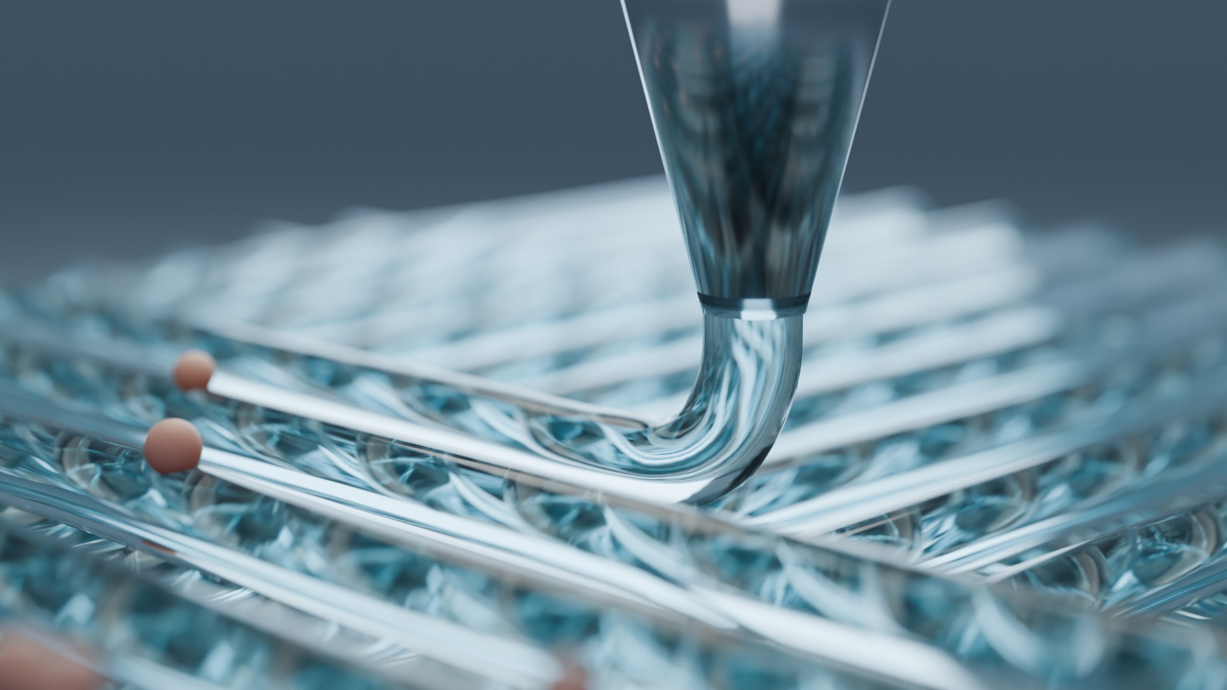
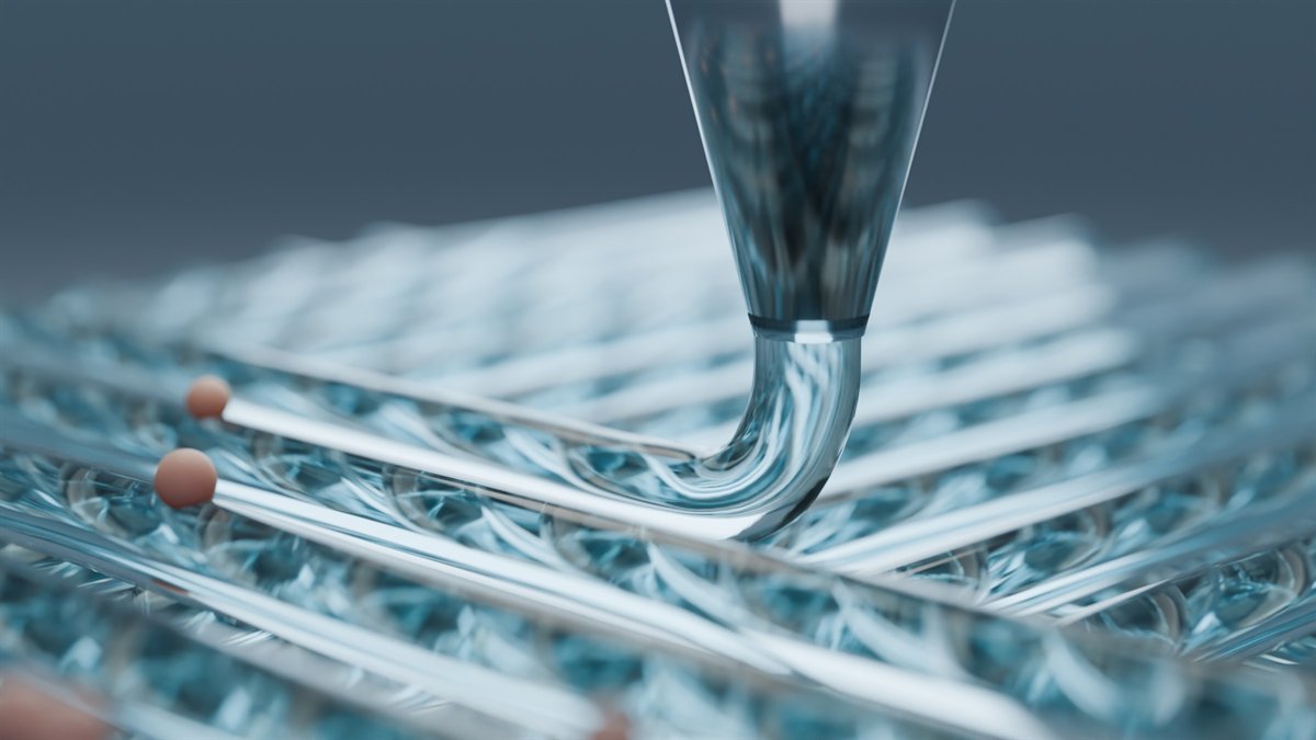
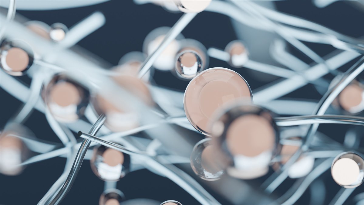
An ink

A cell-friendly hydrogel is pushed through a nozzle as one continuous strand.

Marie Skłodowska-Curie Postdoctoral Fellowship · 2026

A perfusable, bioprinted human liver on a chip

At Friedrich Schiller University Jena I am bioprinting human liver tissue from a silk fibroin–decellularized extracellular matrix (dECM) bioink and perfusing it inside a microfluidic chip. The aim is to rebuild the liver's microarchitecture and keep hepatocytes working long-term, so the chip can be used to study drug metabolism, liver toxicity, and ADME.

The idea, rendered ↗

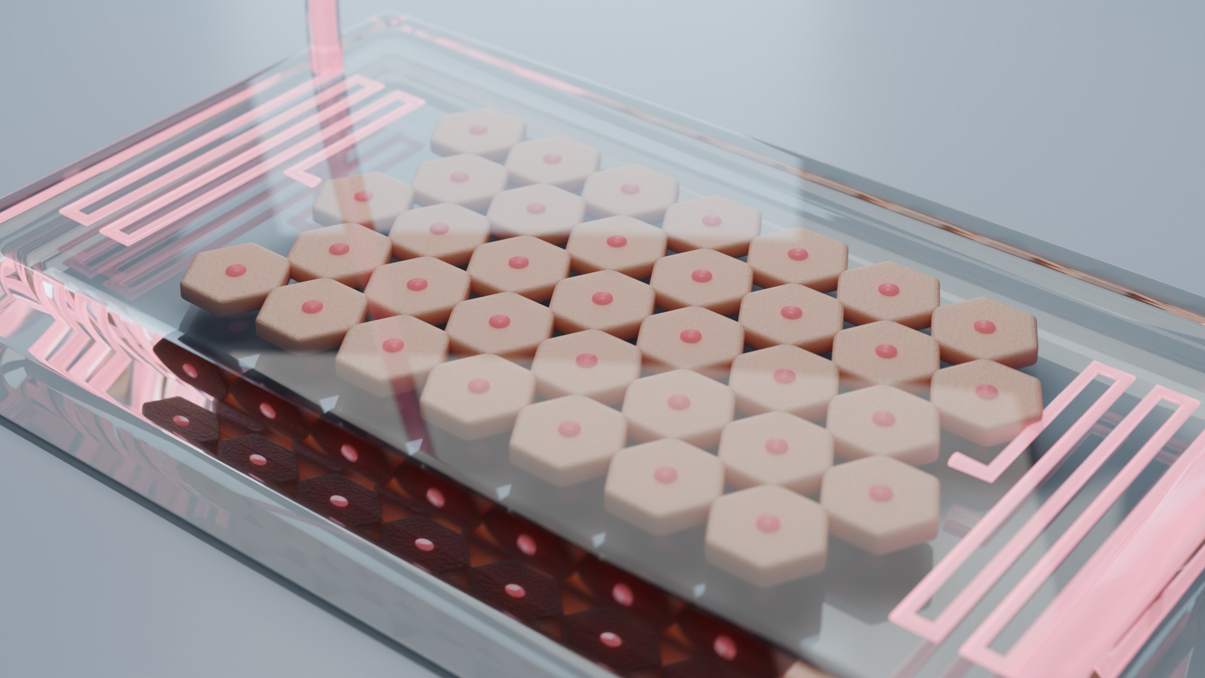
Lobules under flow

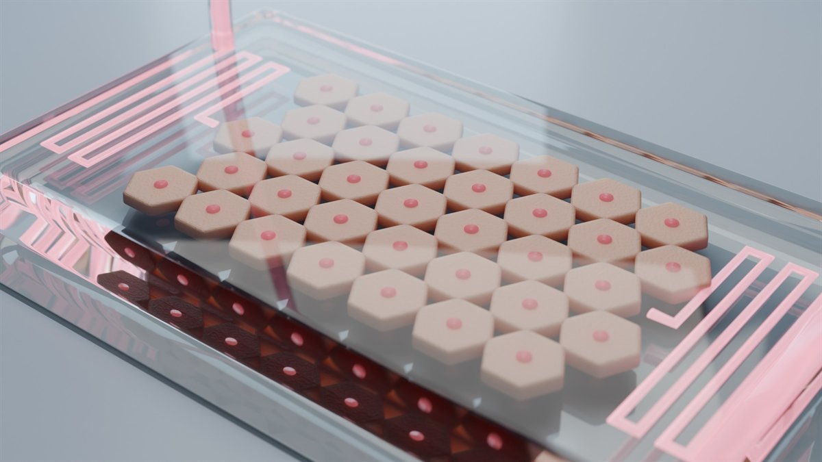
Hexagonal liver lobules printed inside a PDMS chip, fed with culture medium through serpentine channels.

Blender render of a transparent microfluidic chip holding hexagonal printed liver lobules, with pink medium in serpentine channels

A scaffold is a hypothesis about tissue. I make mine specific enough to test: which ink, which stiffness, which signal, and what the cells should do as a result.

My work sits where a printable material meets a living cell. A chitosan–agarose ink loaded with zinc oxide nanoparticles has to kill bacteria and still let stem cells become bone. A conductive chitosan–polyaniline lattice has to keep heart-muscle cells connected and release the exosomes that protect them. A silk–dECM ink has to hold liver cells in the shape of a lobule while medium flows past.

The toolkit stays the same across tissues (extrusion bioprinting, rheology, materials characterization, and cell assays), and so does the rule: every scaffold ends in a number someone else can measure again.

Implant surfaces

TiO₂ nanotubes, drug release

Tehran, 2016–20

Bioink design

Machine-learning-guided printability

Brno, 2020–25

Heart scaffolds

Conductive hydrogels, exosomes

Brno / Northwestern, 2020–25

Bone scaffolds

Nanoparticle-loaded chitosan–agarose

CEITEC, 2023–24

Photosynthetic patches

Chloroplast-integrated hydrogels

Mendel University, 2025–26

Liver-on-a-chip

Silk–dECM bioink under perfusion

FSU Jena, 2026–

Selected work

Peer-reviewed papers, 2020–2026. Each card pairs the result with a Blender scene of the idea the paper tested.

PublishedWound healingPlasma

Plasma-processed bioprinted chitosan hydrogels: enhancing physicochemical and antibacterial properties for wound healing

M. Ezati, N. Mohamad, A. Hashemi, K. Polášková, D. Nečas, I. Zumberg, V. Čmiel, N. Souawda, J. Schäfer, L. Zajíčková (2026). Plasma Processes and Polymers, 23(3), e70151.

What did we find?

Treating the chitosan solvent with a cold atmospheric-pressure plasma jet before printing made the scaffolds stronger, more heat-stable, and more antibacterial, cutting bacterial counts at least 100-fold. Treating the printed scaffolds instead made their surface more bioactive, so cells attached and multiplied better.

Plasma makes printed dressings tougher on bacteria
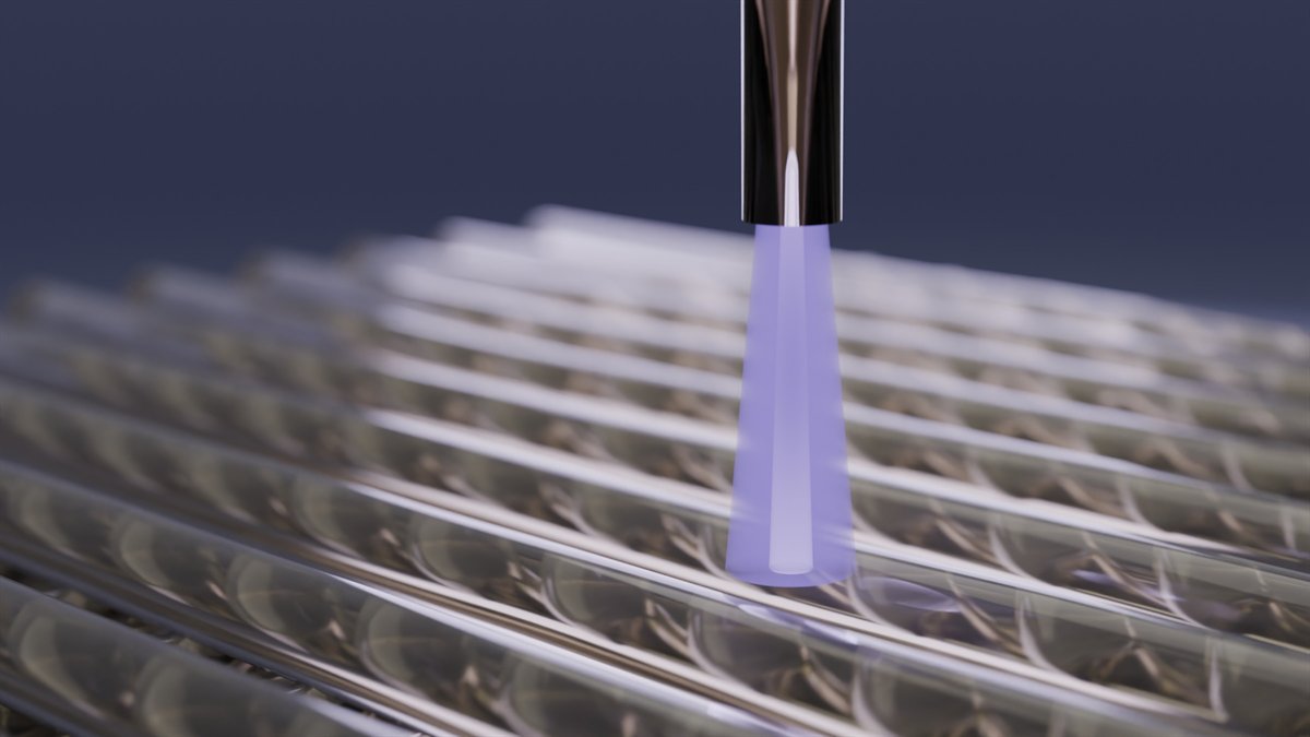
Render of a violet cold-plasma jet over a printed chitosan lattice
A cold-plasma jet glows over a printed chitosan lattice.
PublishedFirst authorBone

Comparative effects of ZnO, MgO, and CaO nanoparticles in 3D-printed chitosan–agarose scaffolds on antibacterial and osteogenic outcomes

A. Hashemi, M. Ezati, R. Paul, I. Zumberg, J. Bacovsky, Z. Fohlerova, V. Provaznik (2025). Macromolecular Bioscience, 25(12), e00232.

What did we find?

A blend of 3.5% chitosan and 1.5% agarose printed best. Of the three nanoparticles, zinc oxide stood out: it raised activity against E. coli and S. aureus and still supported stem-cell growth and bone-type differentiation. Magnesium oxide supported less cell growth, and calcium oxide was the weakest against bacteria.

Zinc oxide wins on both bacteria and bone
Render of a chitosan–agarose lattice studded with three colours of nanoparticles
Three nanoparticle types stud a chitosan–agarose lattice; rod-shaped bacteria sit at its edge.
PublishedFirst authorHeartExosomes

In vitro evaluation of 3D-printed conductive chitosan–polyaniline scaffolds with exosome release for enhanced angiogenesis and cardiomyocyte protection

A. Hashemi, M. Ezati, I. Zumberg, L. Chmelíková, Z. Fohlerová, V. Provazník (2025). RSC Advances, 15(21), 16826–16844.

What did we find?

In a hypoxia–reoxygenation model of heart-attack injury, polyaniline made the printed scaffold conductive, which helped HL-1 heart cells survive and stay connected under low oxygen. Stem-cell exosomes released from the scaffold boosted endothelial migration and tube formation. The two worked together: protected heart cells and more new vessels.

Conduction plus exosomes protects heart cells
Render of heart-muscle cells on a dark conductive lattice with glowing exosomes
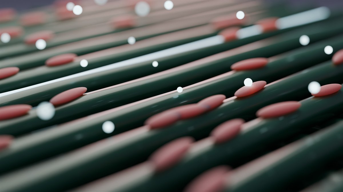
Elongated heart cells line a dark, conductive lattice while exosomes drift above it.
PublishedFirst authorBioinksMachine learning

Characterization and optimization of a biomaterial ink aided by machine learning-assisted parameter suggestion

A. Hashemi, M. Ezati, I. Zumberg, T. Vicar, L. Chmelikova, V. Cmiel, V. Provaznik (2024). Materials Today Communications, 40, 109777.

What did we find?

Machine learning guided the design of a chitosan–gelatin–agarose ink for extrusion bioprinting. The optimized ink had a viscosity suited to reliable printing, printed shapes held, and bone-marrow stem cells grown on it stayed viable. The best formulation was 27% agarose, 53% chitosan, and 20% gelatin.

A model suggests the recipe; the printer checks it
Render of a nozzle printing a new strand onto a hydrogel lattice
The nozzle lays down the next strand of an optimized ink.
ReviewFirst authorVesicles

Extracellular vesicles and hydrogels: an innovative approach to tissue regeneration

A. Hashemi, M. Ezati, M. Partovi Nasr, I. Zumberg, V. Provaznik (2024). ACS Omega, 9(6), 6184–6218.

What does it argue?

Extracellular vesicles carry proteins, lipids, and nucleic acids that can steer cells toward repair, but on their own they are cleared from the body quickly. The review covers where vesicles come from, how they are characterized, and which hydrogel properties let a gel hold them at an injury site for controlled release.

A gel keeps the message where it is needed
Render of glassy vesicles caught in a fibre network
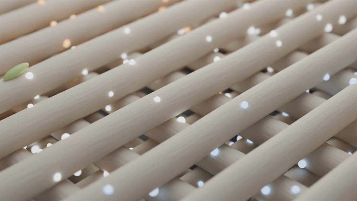
Vesicles, each carrying cargo, caught in a hydrogel fibre network.
PublishedFirst authorImplantsDrug release

Chitosan coating of TiO₂ nanotube arrays for improved metformin release and osteoblast differentiation

A. Hashemi, M. Ezati, J. Mohammadnejad, B. Houshmand, S. Faghihi (2020). International Journal of Nanomedicine, 15, 4471–4481.

What did we find?

Titanium was covered with TiO₂ nanotubes, filled with metformin, and spin-coated with a thin chitosan layer. Compared with uncoated nanotubes, the coated surfaces improved stem-cell attachment, growth, and bone-differentiation markers, and stretched drug release from 3 days to 20 days with a much smaller initial burst.

A thin coat turns 3 days of release into 20
Render of a chitosan film over an array of titanium dioxide nanotubes with glowing drug molecules
A chitosan film drapes over TiO₂ nanotubes; metformin escapes from the open tubes.

All publications

News

Papers, positions, and milestones.

  • PositionStarted as a Marie Skłodowska-Curie Postdoctoral Fellow at the Institute of Pharmacy, Friedrich Schiller University Jena, hosted by Prof. F. Philipp Seib.
  • AwardAwarded a Marie Skłodowska-Curie Postdoctoral Fellowship (Horizon Europe) for a perfusable 3D-bioprinted human liver-on-a-chip with a silk–dECM bioink.
  • PaperPlasma-processed bioprinted chitosan hydrogels for wound healing are out in Plasma Processes and Polymers. Read the paper
  • PaperFirst-author comparison of ZnO, MgO, and CaO nanoparticles in printed bone scaffolds published in Macromolecular Bioscience. Read the paper
  • PaperWith Inna Zumberg and colleagues: local anesthetics ropivacaine and lidocaine curb growth and invasion of osteosarcoma cells, in European Journal of Cell Biology. Read the paper
  • MilestoneDefended the PhD thesis 4D bioprinting based on the maturation of engineered tissue constructs at Brno University of Technology.
  • PaperFirst-author paper on conductive chitosan–polyaniline scaffolds with exosome release published in RSC Advances. Read the paper
  • PositionJoined the Laboratory of intelligent NanoEngineering (iNANO) at Mendel University in Brno to work on a chloroplast-integrated cardiac patch.
Earlier news
  • PaperExosome-loaded chitosan–PEG hydrogels for wound healing, with Masoumeh Ezati, in Macromolecular Bioscience. Read the paper
  • PosterCo-authored poster on bioprinted chitosan hydrogels treated by cold atmospheric plasma jets at ICPM10 & IWPCT9 in Ljubljana.
  • PaperMachine-learning-assisted bioink optimization published in Materials Today Communications. Read the paper
  • ReviewReview on extracellular vesicles and hydrogels for tissue regeneration published in ACS Omega. Read the review
  • PositionResearch scholar at the CEITEC Nano Research Infrastructure, Brno, working on nanoparticle-loaded bone scaffolds.
  • PositionVisiting scholar in Prof. Igor Efimov's laboratory, Department of Biomedical Engineering, Northwestern University, Chicago (until May 2023).
  • TalkOral presentation at the IUPESM World Congress on Medical Physics & Biomedical Engineering (WC2022): machine-learning-based optimization for novel bioink development.
  • PaperChitosan-coated TiO₂ nanotubes for sustained metformin release published in International Journal of Nanomedicine. Read the paper

Seven labs in four countries.

Titanium implant surfaces in Tehran, bioprinted heart scaffolds in Brno, conductive hydrogels in Igor Efimov's lab at Northwestern, nanoparticle bone scaffolds at CEITEC, chloroplast patches at Mendel University, and now a liver-on-a-chip in Jena. Each move added a tool. The habit of ending every project in a measurement stayed the same.

Full CV and background · Google Scholar

9peer-reviewed journal articles, 2019–2026
5as first author, including a review in ACS Omega
208citations on Google Scholar (November 2026)

How the work is done

Every project runs through the same six steps, from a model on screen to a cell response that can be measured.

  1. DesignCAD in Fusion 360; machine-learning parameter suggestion
  2. InkChitosan, agarose, gelatin, PEG, silk–dECM; rheology
  3. PrintExtrusion bioprinting; printability and shape fidelity
  4. CharacterizeSEM, AFM, XRD, XPS, FTIR, Raman; swelling, degradation
  5. CellsMTT, confocal and holographic microscopy, RT-PCR, Western blot
  6. ModelHypoxia–reoxygenation, antibacterial assays, perfusion on chip

Teaching from the same bench

From 2021 to 2024 I taught sessions in Technology in Cellular Engineering (MPA-TBI), a graduate core course at Brno University of Technology: CAD modelling in Autodesk Fusion 360, biomaterials in biomedical engineering, and 3D bioprinting in regenerative medicine. It is the same workflow that sits behind every paper on this site.

Teaching details

Who the work is done with

Long-standing collaborators include Prof. Valentyna Provazník and Prof. Zdenka Fohlerová (Brno University of Technology), Prof. Lenka Zajíčková (plasma processing, Brno), Dr. Xiaohui (Sophie) Ju (Mendel University in Brno), Prof. Igor Efimov (Northwestern University), and Prof. F. Philipp Seib (Friedrich Schiller University Jena), together with co-authors Masoumeh Ezati and Inna Zumberg.

Collaborations · Mentors and co-authors